New landmark imaging research highlights role of thigh muscle size and quality for OA progression
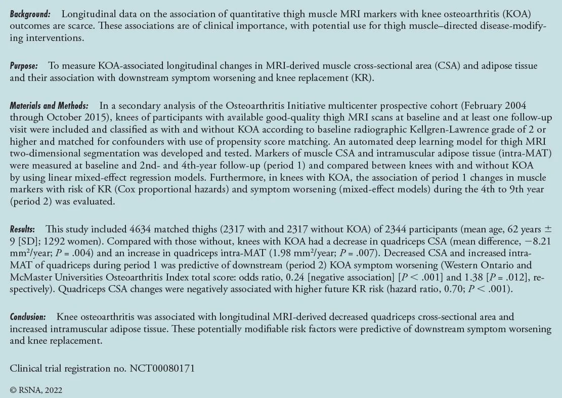
BICL researchers around Drs. Guermazi and Demehri have developed novel methodology to analyze 4634 thigh MRIs from the Osteoarthritis Initiative study. Longitudinal deep learning analysis of thigh MRI scans showed a larger decrease in quadriceps cross-sectional area and increase in quadriceps intramuscular adipose tissue in thighs of knees with baseline osteoarthritis compared to those without. A decrease in quadriceps CSA and increase in intra-MAT were also associated with downstream symptom worsening and knee replacement risk, regardless of osteoarthritis grade.
As previous trials have shown that various interventions can reduce intra-MAT and quadriceps atrophy and increase muscle quality, these results reinforce the role of MRI-derived intra-MAT and quadriceps atrophy as useful markers for the early implementation of secondary preventive measures to improve clinical outcomes in patients with KOA. The results have been published on June 21in the leading journal of medical imaging RADIOLOGY.